What Is It? - Part 2
All of these structures (valves and septums, or septa) are partially formed from Endocardial Cushion tissues. If the Endocardial Cushion tissue fails to develop normally during pregnancy, there may be holes between the heart's chambers, allowing the mixing of blood. The degree of mixing depends on the degrees of tissue development and separation of the heart's chambers.
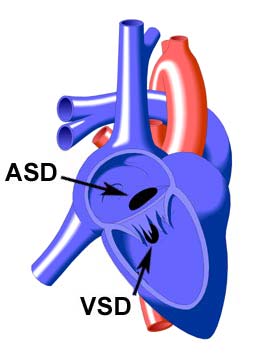
The least complete form of Endocardial Cushion Defect is a hole in the lower Atrial Septum, known as an ostium primum type Atrial Septal Defect (ASD in diagram at left). More extensive defects of this type may involve clefts or deformities in the tricuspid and/or mitral valves, as well as a hole in the upper Ventricular Septum, known as a Ventricular Septal Defect (VSD in diagram at left).
Other names for the more complete forms of Endocardial Cushion Defect are [Atrioventricular Canal Defect (AVC) - Partial], and [Atrioventricular Canal Defect - Complete].
Endocardial Cushion Defect (also known as Atrioventricular Septal Defect) is often associated with the genetic condition known as Down's Syndrome. |